A 20 years old male previously in good health presented with a history of bouts of cough from the last week.
The cough was associated with a low-grade fever but got settled after two days. It was not associated with any specific posture or day-night variation.
The patient denied any history of shortness of breath, orthopnea or paroxysmal nocturnal dyspnea.
He had occasional heartburn but no history of regurgitation or retrosternal chest pain.
Physical examination …
Physical examination was unremarkable. The chest was symmetrical and moving equally with respiration. Breath sounds were vesicular with no added sounds.
Cardiovascular examination revealed a normally placed apex beat with normal cardiac S1 and S2 sounds. No murmur or other added sounds were appreciable.
The abdomen was soft and nontender. No visceromegaly was appreciable.
Investigations of the patient with bouts of cough …
Blood CBC and chemistry was completely normal.
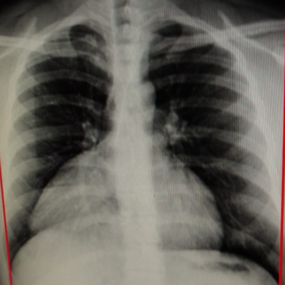
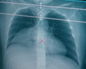
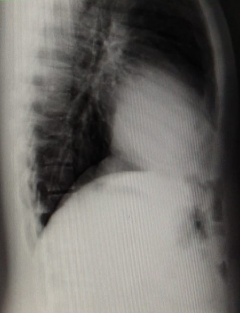
These are his chest radiographs:


What investigations should be advised next?
As most of you think, an Echocardiography should be done next, likewise, we had the same thoughts.
However, Echocardiography and ECG were completely normal.
The next step was to go for a contrast-enhanced CT chest.
Incidental findings pic.twitter.com/PAnRAEmWSQ
— Ahmed Farhan (@DrAhmedFarhan) September 25, 2019
CECT chest was reported as “Morgagni Hernia”
What is a Morgagni hernia?
It is a congenital defect in the diaphragm resulting in herniation of the abdominal contents into the chest.
Most patients are asymptomatic and diagnosed in adulthood.
Rarely, incarceration of the abdominal content may occur and result in severe pain and bleeding.